Differential Scanning Fluorimetry (DSF)
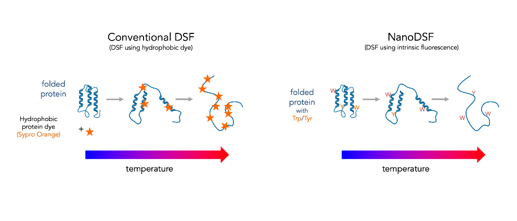
Conventional Differential Scanning Fluorimetry (DSF) uses a real-time PCR instrument to monitor thermally induced protein denaturation by measuring changes in fluorescence of a dye that binds preferentially to unfolded protein (such as Sypro Orange).
Differential Scanning Fluorimetry (DSF)
Conventional Differential Scanning Fluorimetry (DSF) uses a real-time PCR instrument to monitor thermally induced protein denaturation by measuring changes in fluorescence of a dye that binds preferentially to unfolded protein (such as Sypro Orange, which binds to hydrophobic regions of proteins exposed by unfolding). This experiment is also known as a Protein Thermal Shift Assay, because shifts in the apparent melting temperature can be measured upon the addition of stabilizing or destabilizing binding partners or buffer components.
Fluorescent Dyes in DSF
In addition to FITC, Sypro Orange is another widely utilized fluorescent probe in DSF technology, commonly employed in the biomedical field for conducting molecular interaction studies and drug screening. In addition to Sypro Orange, there exist several other fluorescent dyes with similar characteristics that have been demonstrated to be suitable for protein-ligand binding experiment in several studies. For example, Terence L. Kirley's team identified another two fluorescent dyes known as DASPMI (4-(4-(dimethylamino)styryl)-N-methylpyridinium iodide) and Thioflavin T as viable DSF reporters for the analysis of antibody thermal stabilization. By employing these dyes, they accurately calculated the binding affinities of cocaine and its metabolites to h2E2 mAb through quantitative thermodynamic analysis.
Technical Limitations
However, these techniques still share some common drawbacks, such as limitations in detergent or surfactant compatibility and non-selective binding to other hydrophobic reagents. In addition, the additive dyes may also engage in competitive binding with endogenous proteins or hydrophobic ligands, thereby potentially impacting the accuracy of molecular interaction determinations.
New Generation DSF
New generation DSF is a modified differential scanning fluorimetry method which monitors intrinsic tryptophan and tyrosine fluorescence as a function of temperature, time, or denaturant concentration. Tryptophan and tyrosine fluorescence intensity and wavelength maximum will vary as the local chemical environment changes, with significant changes occurring as buried or packed aromatic side chains become solvent exposed upon unfolding. It measures fluorescence intensity at 350 nm and 330 nm and compares the ratio as a function of temperature or denaturant concentration.
Key Advantages
Broader Sample Range
Can be used for a broader range of protein samples than traditional DSF
Higher Throughput
Significantly higher throughput and lower sample consumption than DSC or CD
Comparable Accuracy
Free energies of folding and temperatures of unfolding comparable to DSC values
Label-Free Detection
Monitors intrinsic tryptophan and tyrosine fluorescence without external dyes
Note: The only significant limitation is that the protein of interest must contain aromatic amino acids (tryptophan or tyrosine).
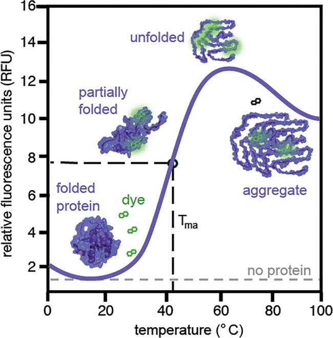
An idealized plot from a differential scanning fluorimetry (DSF) experiment. At low temperature, the target protein is folded and the dye's fluorescence is quenched. As the temperature increases, the protein unfolds, revealing hydrophobic residues. The dye's fluorescence increases when interacting with these regions. In some cases, the fluorescence can decrease at high temperatures, likely because the protein starts to aggregate and exclude dye-binding sites. Importantly, fluorescence is low in the absence of the protein (dotted red line). The half maximal of the fluorescence vs temperature plot is the apparent melting temperature (Tma), which is an indication of the protein's relative stability.