Core Technologies
Advanced analytical technologies for comprehensive protein characterization
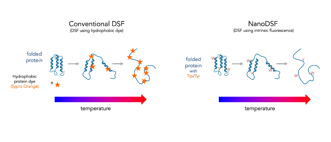
Conventional Differential Scanning Fluorimetry (DSF) uses a real-time PCR instrument to monitor thermally induced protein denaturation by measuring changes in fluorescence of a dye that binds preferentially to unfolded protein (such as Sypro Orange, which binds to hydrophobic regions of proteins exposed by unfolding). This experiment is also known as a Protein Thermal Shift Assay, because shifts in the apparent melting temperature can be measured upon the addition of stabilizing or destabilizing binding partners or buffer components.
ConFix integrates three complementary analytical technologies — Differential Scanning Fluorimetry (DSF), Dynamic Light Scattering (DLS), and Static Light Scattering (SLS) — into a single platform for comprehensive protein characterization.
Fluorescent Dyes in DSF
In addition to FITC, Sypro Orange is another widely utilized fluorescent probe in DSF technology, commonly employed in the biomedical field for conducting molecular interaction studies and drug screening.
Technical Limitations
However, these techniques still share some common drawbacks, such as limitations in detergent or surfactant compatibility and non-selective binding to other hydrophobic reagents.
New Generation DSF
New generation DSF is a modified differential scanning fluorimetry method which monitors intrinsic tryptophan and tyrosine fluorescence as a function of temperature, time, or denaturant concentration.
Key Advantages
Broader Sample Range
Can be used for a broader range of protein samples than traditional DSF
Higher Throughput
Significantly higher throughput and lower sample consumption than DSC or CD
Comparable Accuracy
Free energies of folding and temperatures of unfolding comparable to DSC values
Label-Free Detection
Monitors intrinsic tryptophan and tyrosine fluorescence without external dyes
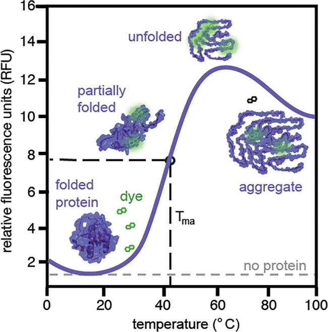
An idealized plot from a differential scanning fluorimetry (DSF) experiment. At low temperature, the target protein is folded and the dye's fluorescence is quenched. As the temperature increases, the protein unfolds, revealing hydrophobic residues. The half maximal of the fluorescence vs temperature plot is the apparent melting temperature (Tma), which is an indication of the protein's relative stability.